Dr. Wang Recent Publications
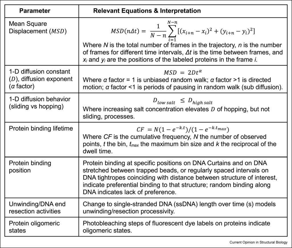
- Single-molecule fluorescence imaging of DNA maintenance protein binding dynamics and activities on extended DNA. EM Irvin, H Wang. Current Opinion in Structural Biology, 87, 102863, 2024.
Abstract: Defining the molecular mechanisms by which genome maintenance proteins dynamically associate with and process DNA is essential to understand the potential avenues by which these stabilizing mechanisms are disrupted. Single-molecule fluorescence imaging (SMFI) of protein dynamics on extended DNA has greatly expanded our ability to accomplish this, as it captures singular biomolecular interactions – in all their complexity and diversity – without relying on ensemble-averaging of bulk protein activity as most traditional biochemical techniques must do. In this review, we discuss how SMFI studies with extended DNA have substantially contributed to genome stability research over the past two years.
- High-speed AFM imaging reveals DNA capture and loop extrusion dynamics by cohesin-NIPBL. Parminder Kaur, Xiaotong Lu, Qi Xu, Elizabeth Marie Irvin, Colette Pappas, Hongshan Zhang, Ilya J Finkelstein, Zhubing Shi, Yizhi Jane Tao, Hongtao Yu, Hong Wang. Journal of Biological Chemistry, 105296, 2023.
Abstract: 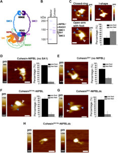 3D chromatin organization plays a critical role in regulating gene expression, DNA replication, recombination, and repair. While initially discovered for its role in sister chromatid cohesion, emerging evidence suggests that the cohesin complex (SMC1, SMC3, RAD21, and SA1/SA2), facilitated by NIPBL, mediates topologically associating domains and chromatin loops through DNA loop extrusion. However, information on how conformational changes of cohesin-NIPBL drive its loading onto DNA, initiation, and growth of DNA loops is still lacking. In this study, high-speed atomic force microscopy imaging reveals that cohesin-NIPBL captures DNA through arm extension, assisted by feet (shorter protrusions), and followed by transfer of DNA to its lower compartment (SMC heads, RAD21, SA1, and NIPBL). While binding at the lower compartment, arm extension leads to the capture of a second DNA segment and the initiation of a DNA loop that is independent of ATP hydrolysis. The feet are likely contributed by the C-terminal domains of SA1 and NIPBL and can transiently bind to DNA to facilitate the loading of the cohesin complex onto DNA. Furthermore, high-speed atomic force microscopy imaging reveals distinct forward and reverse DNA loop extrusion steps by cohesin-NIPBL. These results advance our understanding of cohesin by establishing direct experimental evidence for a multistep DNA-binding mechanism mediated by dynamic protein conformational changes.
- Structure-specific roles for PolG2–DNA complexes in maintenance and replication of mitochondrial DNA. Jessica L Wojtaszek, Kirsten E Hoff, Matthew J Longley, Parminder Kaur, Sara N Andres, Hong Wang, R Scott Williams, William C Copeland. Nucleic Acids Research, gkad679, 2023.
Abstract: 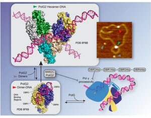 The homodimeric PolG2 accessory subunit of the mitochondrial DNA polymerase gamma (Pol γ) enhances DNA binding and processive DNA synthesis by the PolG catalytic subunit. PolG2 also directly binds DNA, although the underlying molecular basis and functional significance are unknown. Here, data from Atomic Force Microscopy (AFM) and X-ray structures of PolG2-DNA complexes define dimeric and hexameric PolG2 DNA binding modes. Targeted disruption of PolG2 DNA-binding interfaces impairs processive DNA synthesis without diminishing Pol γ subunit affinities. In addition, a structure-specific DNA-binding role for PolG2 oligomers is supported by X-ray structures and AFM showing that oligomeric PolG2 localizes to DNA crossings and targets forked DNA structures resembling the mitochondrial D-loop. Overall, data indicate that PolG2 DNA binding has both PolG-dependent and -independent functions in mitochondrial DNA replication and maintenance, which provide new insight into molecular defects associated with PolG2 disruption in mitochondrial disease.
- Single-molecule Imaging of Genome Maintenance Proteins Encountering Specific DNA Sequences and Structures. EM Irvin, H Wang. DNA Repair, 103528, 2023.
Abstract: 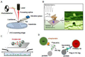 DNA repair pathways are tightly regulated processes that recognize specific hallmarks of DNA damage and coordinate lesion repair through discrete mechanisms, all within the context of a three-dimensional chromatin landscape. Dysregulation or malfunction of any one of the protein constituents in these pathways can contribute to aging and a variety of diseases. While the collective action of these many proteins is what drives DNA repair on the organismal scale, it is the interactions between individual proteins and DNA that facilitate each step of these pathways. In much the same way that ensemble biochemical techniques have characterized the various steps of DNA repair pathways, single-molecule imaging (SMI) approaches zoom in further, characterizing the individual protein-DNA interactions that compose each pathway step. SMI techniques offer the high resolving power needed to characterize the molecular structure and functional dynamics of individual biological interactions on the nanoscale. In this review, we highlight how our lab has used SMI techniques – traditional atomic force microscopy (AFM) imaging in air, high-speed AFM (HS-AFM) in liquids, and the DNA tightrope assay – over the past decade to study protein-nucleic acid interactions involved in DNA repair, mitochondrial DNA replication, and telomere maintenance. We discuss how DNA substrates containing specific DNA sequences or structures that emulate DNA repair intermediates or telomeres were generated and validated. For each highlighted project, we discuss novel findings made possible by the spatial and temporal resolution offered by these SMI techniques and unique DNA substrates.
- Assembly path dependence of telomeric DNA compaction by TRF1, TIN2, and SA1. Ming Liu, Hai Pan, Parminder Kaur, Lucia J Wang, Miao Jin, Ariana C Detwiler, Patricia L Opresko, Yizhi Jane Tao, Hong Wang, Robert Riehn. Biophysical Journal, 122 (10), 1822-1832, 2023.
Abstract: 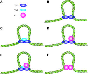 Telomeres, complexes of DNA and proteins, protect ends of linear chromosomes. In humans, the two shelterin proteins TRF1 and TIN2, along with cohesin subunit SA1, were proposed to mediate telomere cohesion. Although the ability of the TRF1-TIN2 and TRF1-SA1 systems to compact telomeric DNA by DNA-DNA bridging has been reported, the function of the full ternary TRF1-TIN2-SA1 system has not been explored in detail. Here, we quantify the compaction of nanochannel-stretched DNA by the ternary system, as well as its constituents, and obtain estimates of the relative impact of its constituents and their interactions. We find that TRF1, TIN2, and SA1 work synergistically to cause a compaction of the DNA substrate, and that maximal compaction occurs if all three proteins are present. By altering the sequence with which DNA substrates are exposed to proteins, we establish that compaction by TRF1 and TIN2 can proceed through binding of TRF1 to DNA, followed by compaction as TIN2 recognizes the previously bound TRF1. We further establish that SA1 alone can also lead to a compaction, and that compaction in a combined system of all three proteins can be understood as an additive effect of TRF1-TIN2 and SA1-based compaction. Atomic force microscopy of intermolecular aggregation confirms that a combination of TRF1, TIN2, and SA1 together drive strong intermolecular aggregation as it would be required during chromosome cohesion.
- PARP1 associates with R-loops to promote their resolution and genome stability. Natalie Laspata, Parminder Kaur, Sofiane Yacine Mersaoui, Daniela Muoio, Zhiyan Silvia Liu, Maxwell Henry Bannister, Hai Dang Nguyen, Caroline Curry, John M Pascal, Guy G Poirier, Hong Wang, Jean-Yves Masson, Elise Fouquerel. Nucleic Acids Research, gkad066, 2023.
Abstract: 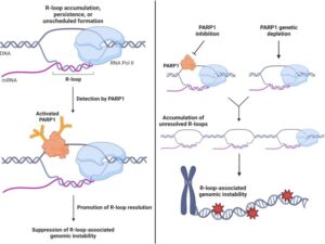 PARP1 is a DNA-dependent ADP-Ribose transferase with ADP-ribosylation activity that is triggered by DNA breaks and non-B DNA structures to mediate their resolution. PARP1 was also recently identified as a component of the R-loop-associated protein-protein interaction network, suggesting a potential role for PARP1 in resolving this structure. R-loops are three-stranded nucleic acid structures that consist of a RNA-DNA hybrid and a displaced non-template DNA strand. R-loops are involved in crucial physiological processes but can also be a source of genome instability if persistently unresolved. In this study, we demonstrate that PARP1 binds R-loops in vitro and associates with R-loop formation sites in cells which activates its ADP-ribosylation activity. Conversely, PARP1 inhibition or genetic depletion causes an accumulation of unresolved R-loops which promotes genomic instability. Our study reveals that PARP1 is a novel sensor for R-loops and highlights that PARP1 is a suppressor of R-loop-associated genomic instability.
